Cataract Surgery Patient Story
Ophthalmology

Sadie, the adorable and spunky Miniature Schnauzer, was referred to Dr. Alexandra Ng by Fisher Animal Hospital for evaluation of cataracts.
Most cataracts are inherited in dogs, but in Sadie's case, it is secondary to diabetes mellitus.
"Fun" fact about diabetic cataracts: About 75-80% of diabetic dogs will develop cataracts within the first year of their diagnosis, regardless of how well-controlled their diabetes is. The cataracts tend to form quickly and can cause severe inflammation inside the eyes. This can lead to permanently blinding and potentially painful eye conditions, such as glaucoma.
Another "fun" fact: Not all cataracts are ideal for cataract surgery. Initial examination and pre-surgical diagnostics are needed to ensure the best chance of restoring sight.
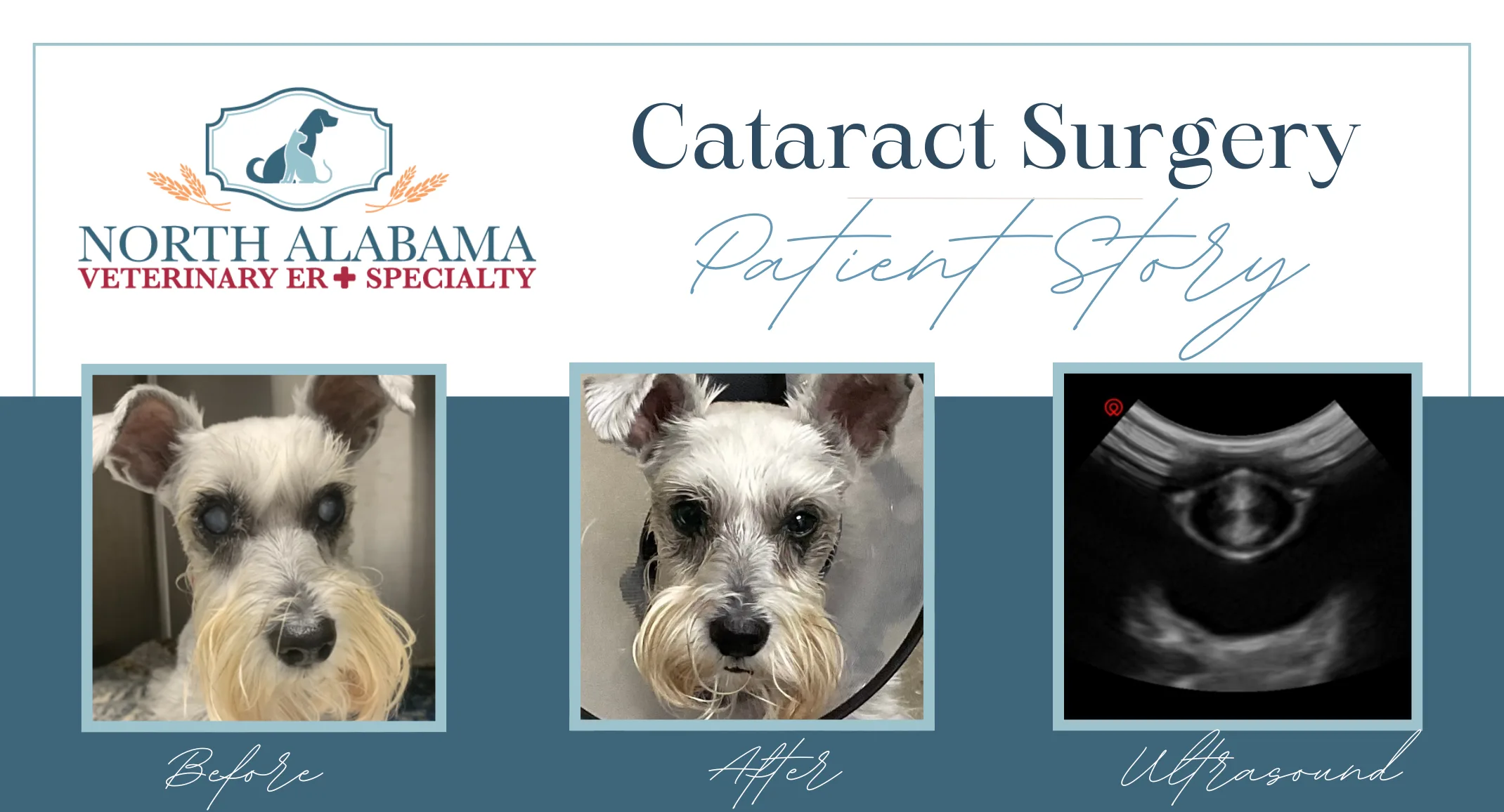
Sadie's initial examination confirmed cataracts that were causing her significant visual impairment. Sadie passed her pre-surgical diagnostics with flying colors and was scheduled for cataract surgery! Diagnostics included ocular ultrasound, showing no evidence of retinal detachment in either eye, and Electroretinogram (ERG), showing normal retinal function in both eyes.
Cataract removal surgery (phacoemulsification) involves making a small incision in the cornea (clear part of the eye) and using handheld equipment to fragment and remove the cataract. An artificial lens is inserted in most cases pending the stability of the lens capsule. Sadie's surgery went well and she received artificial lenses in both eyes.
Post-operative care is intensive and important for surgical success. It includes an Elizabethan collar (cone), multiple eye medications, oral medications, and rest.
Sadie is back to watching the dogs and squirrels outside and loves having her vision back!
